RONAVIS-PN
Real-time navigation and guidance system for fracture reduction surgery
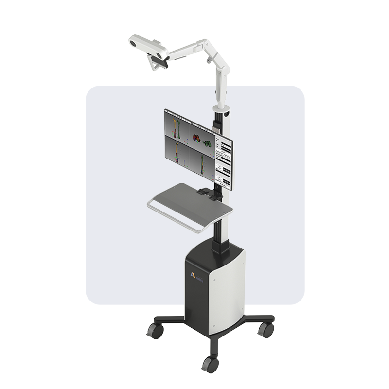
Conventional fracture reduction relies on 2D radiological images for visualisation and assessment – but fractures are three-dimensional injuries. With RONAVIS-PN, orthopaedic surgeons can now better visualise fractures with fewer X-ray exposures, then plan an optimised reduction path and monitor the reduction procedure in real time.

3D visualisation
Using 2D radiological images and external guides, RONAVIS-PN generates 3D models of the patient’s bone fragments to give the surgical team superior visualisation over traditional workflows.
Real-time navigation
Based on the 3D model generated by RONAVIS-PN, surgeons can monitor the positions of bone fragments in real time during fracture reduction. This eliminates the need for repeated imaging to maintain the surgical team’s situational awareness, with up to 90% fewer X-rays required.
Surgical pathfinding
Based on the target endpoint, RONAVIS-PN plans and directs the reduction paths for the bone fragments, which minimise any risks of further injuries to the surrounding tissues.